HRCT Chest vs CT Chest Scan: When Each Lung Scan Is Used
Book Your HRCT Now at Affordable Prices
Persistent cough, breathing difficulty, chest pain, lung infection, or abnormal chest X-ray findings often lead doctors to recommend advanced chest imaging. HRCT chest and standard CT chest scans are different tests designed for different diagnostic needs. One focuses deeply on lung tissue detail, while the other provides broader chest evaluation. Read on to know which scan may suit different lung conditions and why doctors choose one over the other.
Why Doctors Recommend Advanced Chest Imaging
Advanced pulmonary imaging helps doctors identify lung abnormalities, airway disease, infection spread, and structural chest changes that may not appear clearly on regular X rays.
Conditions That Often Lead to Chest Scans
• Persistent cough lasting several weeks without clear improvement
• Breathlessness during walking, climbing, or routine daily activities
• Wheezing sounds suggesting airway narrowing or respiratory inflammation
• Chest tightness causing discomfort during breathing or movement
• Long-term smoking history increasing chronic lung disease risks
• Abnormal chest X ray requiring detailed respiratory disease diagnosis
• Fever with suspected lung infection needing chest imaging test
What HRCT and CT Chest Scans Actually Show
• Lung tissues showing inflammation, fibrosis, or structural abnormalities
• Airways and bronchi revealing blockage, swelling, or airway disease
• Blood vessels identifying clots, narrowing, or vascular complications clearly
• Pleura showing fluid buildup or surrounding lung membrane changes
• Nodules and masses needing detailed pulmonary imaging for evaluation
• Fibrosis patterns linked with chronic diffuse lung disease progression
• Infection spread affecting multiple lung areas during severe pneumonia
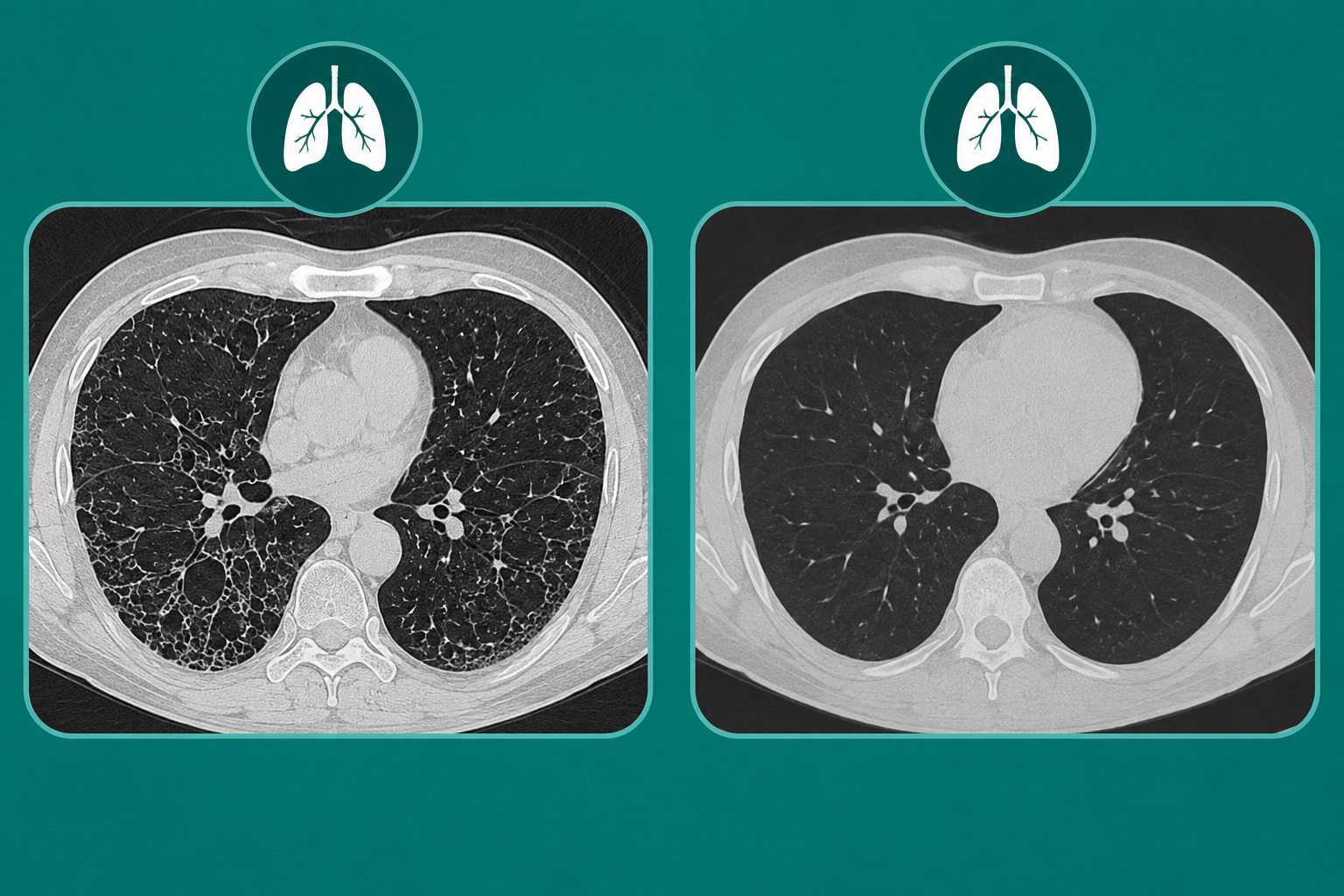
HRCT Chest vs CT Scan: Differences Explained
HRCT chest scan and standard CT chest scans use similar imaging technology, but their diagnostic purpose differs significantly. A pulmonologist or radiologist selects the scan depending on symptoms, suspected disease, and the level of detail required for accurate diagnosis.
| Feature | HRCT Chest Scan | Standard CT Chest Scan |
|---|---|---|
| Image detail | Extremely detailed lung tissue imaging | Broader chest structure imaging |
| Slice thickness | Very thin image slices | Thicker image slices commonly used |
| Main purpose | Evaluates fine lung tissue abnormalities | Evaluates chest organs and vessels |
| Conditions detected | Pulmonary fibrosis, bronchiectasis, COPD | Tumors, trauma, infections, clots |
| Use of contrast | Usually performed without contrast dye | Often performed with contrast |
| Scan duration | Usually shorter focused imaging process | Slightly longer with contrast usage |
| Radiation exposure | Focused lung imaging exposure | Depends on scan protocol selected |
| Lung tissue evaluation | Highly detailed interstitial lung disease assessment | General chest tissue evaluation |
| Emergency usage | Less common during emergencies | Common emergency lung CT scan |
HRCT is not simply a better CT scan. It is specially optimized for detecting fine lung tissue changes linked with pulmonary fibrosis, interstitial lung disease, bronchiectasis, pneumonia, and COPD related structural damage. Standard CT chest scans provide wider chest evaluation and are commonly used for lung nodules, tumors, trauma, blood vessel assessment, and emergency conditions.
Lung Conditions Where HRCT Is Commonly Preferred
HRCT chest scan provides high resolution lung imaging that helps doctors detect subtle tissue abnormalities, chronic respiratory disease imaging patterns, and diffuse lung disease changes more clearly than routine chest CT scans.
Chronic Lung Diseases Seen Clearly on HRCT
HRCT chest scan clearly detects interstitial lung disease, pulmonary fibrosis, bronchiectasis, and COPD related structural changes inside the lungs. It also helps evaluate occupational lung disease caused by dust, chemical exposure, or long term environmental inhalation affecting delicate lung tissues.
Situations Where HRCT Helps Track Disease Progression
Doctors may recommend HRCT for long standing cough, autoimmune lung involvement, post infection lung scarring, or follow up after severe pneumonia. It also helps monitor treatment response in fibrotic lung disease and chronic inflammatory respiratory conditions affecting breathing capacity over time.
When a Standard CT Chest Scan Is Usually Recommended
A standard CT chest with contrast is commonly chosen when doctors need broader chest evaluation involving blood vessels, tumors, infection complications, or emergency conditions. It helps assess multiple chest structures quickly and accurately.
• Chest trauma needing rapid internal injury assessment and evaluation
• Lung tumors and masses requiring detailed structural chest imaging
• Cancer staging checking spread into surrounding chest structures carefully
• Pulmonary embolism evaluation detecting dangerous blood clots within vessels
• Severe infection complications affecting multiple chest regions and tissues
• Emergency chest assessment during sudden breathing or chest problems
• Mediastinal evaluation assessing central chest organs and lymph nodes
• Pleural effusion assessment detecting abnormal fluid around lung lining
Contrast enhanced CT scan for chest infection, vascular disease, or chest CT for cancer detection helps visualize blood vessels, tumors, and chest organs more clearly. HRCT mainly focuses on fine lung tissue detail rather than broader vascular and emergency chest evaluation.
Preparing for the Scan and What Happens During the Procedure
Both HRCT chest and CT chest scans are simple imaging procedures completed within minutes. Preparation varies slightly depending on whether contrast dye is required during the scan.
• Whether fasting is needed: Fasting may be advised before CT chest with contrast, especially when contrast dye injection is planned for vascular or tumor evaluation.
• Contrast dye instructions: Inform doctors about allergies, kidney disease, or previous contrast reactions before undergoing contrast enhanced chest imaging procedures safely.
• Clothing and metal objects: Comfortable clothing without metallic objects helps avoid imaging interference during pulmonary imaging and chest scan procedures inside radiology departments.
• Scan duration: Most HRCT chest and CT chest scans finish within ten to twenty minutes, depending on scan complexity and contrast usage.
• Breathing instructions: Patients may briefly hold breath during scanning for clearer lung tissue images and accurate respiratory disease diagnosis results.
• Safety considerations in pregnancy: Pregnant women should inform healthcare providers before chest imaging because radiation exposure requires careful medical evaluation during pregnancy.
• When reports are typically reviewed by a radiologist: Experienced radiologists usually review scan findings within hours or a few days depending on urgency and hospital workflow.
Choosing the Right Scan for Better Lung Evaluation
Choosing between HRCT chest and CT chest scan depends on symptoms, suspected disease, and the level of detail doctors require. HRCT is highly useful for chronic lung tissue evaluation, while standard CT chest scans help assess tumors, trauma, blood vessels, and emergency chest conditions. A pulmonologist and radiologist together decide the most suitable chest imaging approach for accurate diagnosis, treatment planning, and long term respiratory care.
Safe and Affordable HRCT Chest Scan in Dubai
Amax Healthcare provides advanced HRCT chest scan and CT chest imaging by connecting patients with trusted diagnostic centers and experienced radiologists across Dubai. Our network supports safe, accurate, and affordable lung imaging for respiratory disease diagnosis, pulmonary fibrosis evaluation, chest infection assessment, and chronic airway disease monitoring. We focus on quick appointments, reliable reporting, experienced specialists, and patient friendly diagnostic support for better respiratory healthcare management.
FAQs
Is HRCT chest more accurate than a normal CT scan for lung disease?
HRCT chest is more accurate for lung disease because it shows detailed lung tissue changes linked with fibrosis, bronchiectasis, airway disease, and chronic respiratory conditions.
Why would a doctor order HRCT instead of a CT chest scan?
A doctor orders HRCT instead of CT chest scan when detailed lung tissue evaluation is needed for interstitial lung disease, pulmonary fibrosis, or chronic respiratory symptoms.
Can HRCT detect lung infection and pneumonia?
HRCT can detect lung infection and pneumonia by showing infection spread, inflammation, lung scarring, and structural respiratory changes more clearly than standard imaging.
Does HRCT chest use contrast dye?
HRCT chest usually does not use contrast dye because the scan mainly focuses on detailed lung tissue imaging rather than blood vessels or tumors.
Which scan is better for pulmonary fibrosis and lung scarring?
HRCT is better for pulmonary fibrosis and lung scarring because it provides high resolution lung imaging with detailed fibrotic tissue evaluation.
Is there more radiation exposure in HRCT compared to CT chest?
Radiation exposure in HRCT compared to CT chest depends on scan protocol, slice thickness, and imaging requirements selected by the radiologist.
Can HRCT and CT chest detect lung cancer?
HRCT and CT chest can detect lung cancer, but standard CT chest with contrast is commonly preferred for tumor evaluation and cancer staging.