

Early Nerve Compression Detection with 3T Neuro MRI Scans
Nerve compression often begins quietly. The pain may feel mild, the numbness may come and go, and many people delay testing because symptoms do not seem serious. Unfortunately, early nerve pressure can slowly cause lasting damage if it is not identified on time. A 3T neuro MRI scan helps doctors see nerves more clearly and detect compression at an early stage. This blog explains how neuro MRI scans work, why 3T MRI is important, and how early detection improves outcomes.
Early Nerve Compression
Nerve compression happens when a nerve is pressed by surrounding structures. This pressure may come from a disc, bone, muscle, ligament, or swelling. When the pressure is mild, symptoms may not be constant. This makes early nerve compression difficult to recognize.
In the early phase, nerves may still function, but they become irritated. Over time, ongoing pressure reduces blood supply to the nerve and affects signal transmission. If this continues, the nerve can weaken or lose function permanently.
Early nerve compression commonly affects areas such as the spine, neck, and brain related nerve pathways. Neuro MRI scans help doctors see these areas in detail and understand where the pressure is happening.
Common Symptoms That Point to Nerve Compression
Symptoms of nerve compression vary depending on the nerve involved. Some people feel pain, while others notice weakness or loss of sensation. Early symptoms are often ignored because they may improve temporarily.
Common signs include:
- Tingling or pins and needles sensation
- Numbness in arms, legs, hands, or feet
- Localized pain that spreads along a nerve path
- Muscle weakness or reduced grip strength
- Burning or electric-like sensations
These symptoms may worsen with certain movements or positions. They may also appear only during activity or at night. Neuro MRI scans help identify the cause when symptoms are unclear.
Why MRI Is the Preferred Scan for Nerve Imaging
How MRI Visualizes Nerves and Soft Tissues
MRI uses magnetic fields and radio waves to create detailed images of soft tissues. Unlike X-rays or CT scans, MRI does not rely on radiation. This makes it ideal for viewing nerves, discs, muscles, and spinal cord structures.
Neuro MRI scans show:
- Nerve roots and spinal cord
- Discs and surrounding soft tissue
- Brain structures and cranial nerves
Because nerves are soft tissues, MRI provides better contrast and clarity than other imaging methods.
Limitations of Standard MRI in Early Detection
Standard MRI scanners may not always detect subtle nerve compression. Lower resolution images can miss early changes such as mild swelling or slight disc bulging.
Limitations may include:
- Reduced image detail
- Difficulty separating nerves from nearby tissue
- Motion-related image blur
This is where 3T neuro MRI scans offer a clear advantage.
What Makes 3T Neuro MRI Different
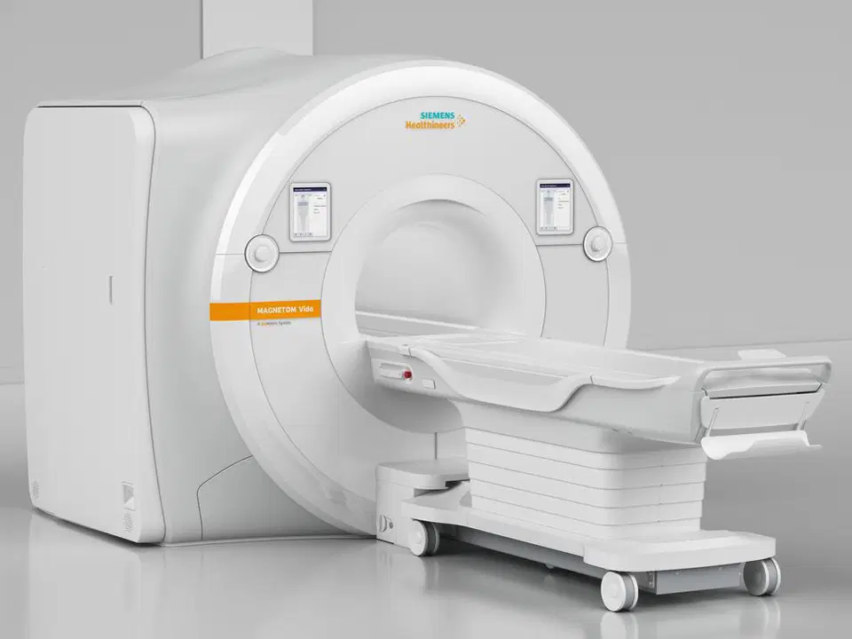
A 3T MRI scanner uses a stronger magnetic field compared to standard systems. The stronger field produces clearer and more detailed images.
The main differences include:
- Higher image resolution
- Better contrast between nerves and tissues
- Improved ability to detect small changes
3T neuro MRI scans allow doctors to see fine nerve structures that may not appear clearly on standard scans. This makes early nerve compression easier to identify.
How 3T Neuro MRI Detects Nerve Compression Earlier
Higher Resolution and Clearer Nerve Detail
3T neuro MRI scans provide sharper images of nerve roots and spinal cord pathways. Even mild compression becomes visible because the boundaries between nerves and surrounding tissue are clearer.
This helps doctors:
- Identify exact compression points
- Assess nerve swelling or irritation
- Detect early disc-related pressure
Early identification allows treatment to begin before symptoms worsen.
Improved Signal and Image Accuracy
The stronger magnetic field improves signal strength. This reduces background noise and improves image accuracy.
Benefits include:
- Clear separation of nerves and discs
- Better visualization of spinal canal space
- More reliable diagnosis
This accuracy reduces the chance of missed findings and supports confident clinical decisions.
Areas Commonly Evaluated in Neuro MRI Scans
Neuro MRI scans focus on areas where nerves are commonly affected. These scans help doctors understand both structure and nerve condition.
Common areas include:
- Brain and cranial nerves
- Neck and cervical spine
- Thoracic spine
- Lumbar spine
- Peripheral nerve pathways when required
Each area is evaluated carefully to locate compression and assess its severity.
Who Benefits Most from a 3T Neuro MRI Scan
Many people benefit from 3T neuro MRI scans, especially when symptoms are unclear or persistent.
This scan is often useful for:
- People with ongoing pain or numbness
- Individuals whose earlier scans were normal but symptoms continue
- Athletes with nerve-related injuries
- Patients with nerve symptoms after surgery
- Individuals with suspected spinal or neurological conditions
Early imaging helps guide treatment and reduces uncertainty.
When Doctors Recommend 3T Neuro MRI Over Standard MRI
Doctors may suggest a 3T neuro MRI scan when standard imaging does not provide enough information. This decision is based on symptoms, medical history, and clinical examination.
Common reasons include:
- Persistent symptoms with unclear diagnosis
- Planning precise treatment or intervention
- Monitoring nerve-related conditions over time
- Evaluating complex spinal or brain nerve issues
Choosing the right imaging method improves diagnostic confidence.
What to Expect During a 3T Neuro MRI Scan
Understanding the scan process helps reduce anxiety. A 3T neuro MRI scan is safe and noninvasive.
During the scan:
- You lie still on the scanner table
- The scan takes around thirty to sixty minutes
- You may hear loud sounds, but ear protection is provided
- Contrast may be used if required
After the scan, normal activities can usually resume. Results are reviewed by experienced radiology specialists.
Accuracy and Safety of 3T Neuro MRI Scans
Diagnostic Accuracy for Nerve Compression
3T neuro MRI scans offer high diagnostic accuracy. Clear images allow early detection of nerve pressure before major damage occurs.
Accurate diagnosis helps:
- Reduce delays in treatment
- Avoid unnecessary procedures
- Support targeted therapy planning
Early detection often leads to better recovery.
Safety Considerations
MRI scans do not use radiation. This makes them safe for repeated use when needed.
Safety features include:
- Standard screening for metal implants
- Careful monitoring during scanning
- Use of approved contrast agents when required
Most patients tolerate 3T neuro MRI scans well.
How Early Detection Changes Treatment Outcomes
Early nerve compression detection improves treatment success. When compression is identified early, treatment options are often simpler and more effective.
Benefits include:
- Better response to physical therapy
- Reduced need for invasive procedures
- Lower risk of permanent nerve damage
- Faster symptom relief
Early imaging helps doctors choose the right approach and monitor progress accurately.
Frequently Asked Questions
What is a neuro MRI scan used for
A neuro MRI scan is used to evaluate the brain, spine, and nerves. It helps diagnose nerve compression, inflammation, and structural problems.
Is 3T MRI better than standard MRI for nerve problems
Yes. A 3T MRI provides clearer images and better nerve detail. This improves early detection and diagnostic accuracy.
Can a neuro MRI detect nerve compression early
Yes. Neuro MRI scans, especially with 3T technology, can identify early nerve pressure before severe symptoms develop.
When should I choose a 3T neuro MRI scan
A 3T neuro MRI scan is recommended when symptoms persist, earlier scans are unclear, or precise diagnosis is required.
Is 3T MRI safe for repeated scans
Yes. MRI does not use radiation and is considered safe for repeated imaging when medically necessary.
Choosing 3T Neuro MRI Early Improves Long-Term Nerve Health
Early nerve compression detection plays a critical role in preventing long-term nerve damage. A 3T neuro MRI scan offers clearer images, higher accuracy, and better confidence in diagnosis. By identifying nerve pressure early, doctors can guide treatment effectively and reduce the risk of lasting symptoms. Choosing advanced neuro MRI imaging supports timely care, better recovery, and improved quality of life.
If you are looking for a trusted 3T neuro MRI in Dubai, Amax connects you with advanced imaging centers equipped with high-resolution 3T MRI systems. Through experienced radiology experts and clear guidance, Amax helps ensure accurate neuro MRI scans for early nerve compression detection at affordable and transparent pricing.